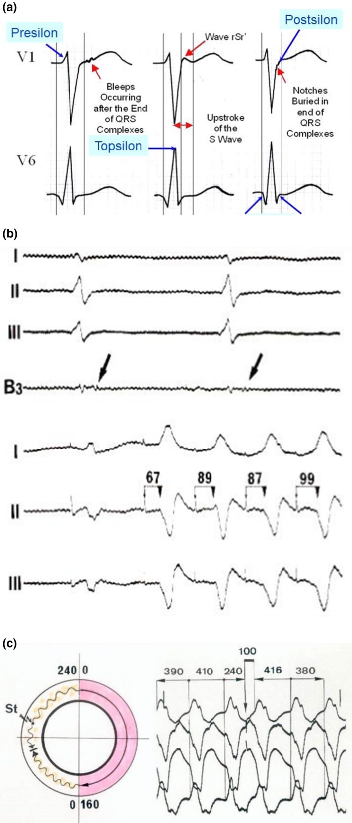
Figure 4.

(a) The new names for identification of depolarization abnormalities (The original Epsilon wave was recorded on the epicardium with an interelectrode distance of 1.5 mm) (With permission from Dr Guoliang Li (Li et al., 2018)). (b) Example of stimulation in the zone of slow conduction of arrhythmogenic right ventricular dysplasia (ARVD). Upper tracings: Recording of late potentials on the epicardium. The stimulation in the same zone shows a delay between the stimulus artifact and the ventricular activation on the surface leads. This was possible because all electrophysiological parameters were stored on a magnetic tape during each procedure. Several years later, this phenomenon led to the concept of concealed entrainment presented on the tracing below. Entrainment by stimulation in the zone of slow conduction is obtained after a single stimulus. (c) An excellent demonstration of concealed entrainment with a single stimulus delivered in the zone of slow conduction. Note the typical ECG pattern of VT with LBBB pattern and superior axis in a patient with typical ARVD. There is a delay of 100 ms in between the stimulus and the ventricular response. Note that the morphology of the “entrained QRS” is exactly the same that the morphology observed during spontaneous VT and that this morphology is also followed by the same morphology with the same coupling beat to beat interval of 380–390 ms (With permission from Dr. Guy Fontaine (Fontaine, 2010))
