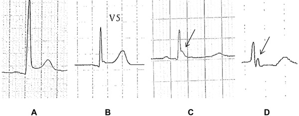
Figure 3.

Different early repolarization patterns with different ST segment shifts: upsloping ST segment elevation (A), horizontal ST segment elevation (B), isoelectric ST segment (C), and ischemic ST segment depression (D). In (C) and (D) the diagnosis of early repolarization is based only on the presence of J waves as indicated by arrows (patients 4, 8, 30, and 42, respectively, among the case group).
