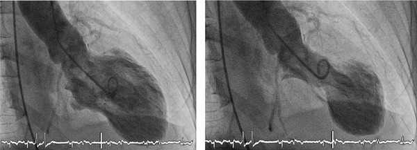
Figure 1.

Example of Takotsubo syndrome ventriculography. Left picture shows left ventricle in diastole. Right picture shows the left ventricle in systole, with the typical shape due to apical dyskinesia and hypercontractility of the basal segments. See electrocardiogram monitoring.
