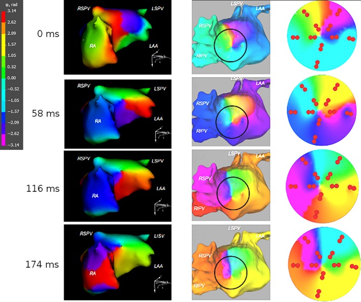
Figure 3.

Noninvasively and invasively imaged rotors during atrial fibrillation (AF). The phase maps obtained by novel noninvasive epicardial and endocardial electrophysiology system (NEEES) (left‐hand panels) and catheter mapping (middle and right‐hand panels) demonstrated a rotor in a similar atrial location rotating in the same clockwise direction
