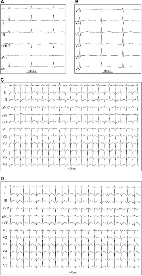
Figure 2.

Representative ECG registrations 15 minutes after flecainide infusion (A) resting ECG of the same patient, as in Fig. 1, 15 minutes after an intravenous infusion of flecainide (2 mg/kg) showing sinus rhythm at rest (limb leads, paper speed 50 mm/s) (B) chest leads (C) ECG during the 7th minute of exercise at a workload of 120 W, demonstrating the absence of PVCs (25 mm/s) (D) resting ECG 1‐minute after cessation of exercise (25 mm/s).
