
Figure 1.
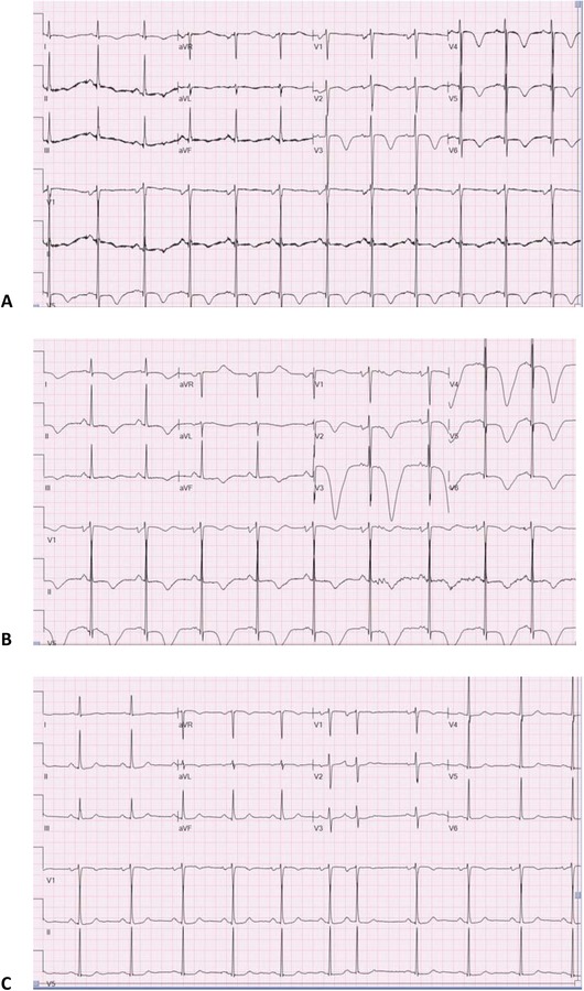
Representative ECG in an AA patient: (A) ECG on admission showing sinus rhythm with diffuse T‐wave inversions, (B) ECG at 48 hours with deeper T‐wave inversions, and (C) ECG at follow‐up with resolution of T‐wave inversions.
Official websites use .gov
A
.gov website belongs to an official
government organization in the United States.
Secure .gov websites use HTTPS
A lock (
) or https:// means you've safely
connected to the .gov website. Share sensitive
information only on official, secure websites.
Representative ECG in an AA patient: (A) ECG on admission showing sinus rhythm with diffuse T‐wave inversions, (B) ECG at 48 hours with deeper T‐wave inversions, and (C) ECG at follow‐up with resolution of T‐wave inversions.