
Figure 2.
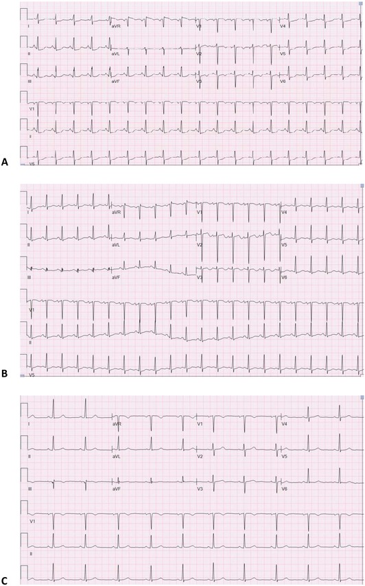
Representative ECG in a non‐AA patient: (A) ECG on admission showing sinus tachycardia with diffuse ST depressions and T‐wave inversions in V1–V3, (B) ECG at 48 hours with persistent T‐wave inversions in V1–V3 and mild improvement of diffuse ST depressions, and (C) ECG at follow‐up with resolution of ST‐depressions and T‐wave inversions.