Figure 1.

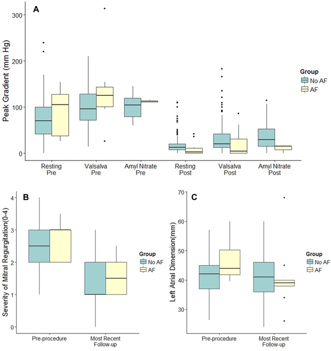
(A) Peak left ventricular outflow tract gradients at baseline and postprocedure in hypertrophic cardiomyopathy patients with and without incident atrial fibrillation (yellow and green fill respectively). Graphical depictions of inter‐quartile range (IQR, 25th–75th percentiles; box), median (horizontal line), and outliers (>1.5*IQR; points). Between groups comparisons were not statistically significant with the exception of with amyl nitrate (0.047 and 0.014 pre = and post = respectively). (B) Severity of Mitral regurgitation on a semi‐quantitative scale of 0–4 (none = 0, trace = 1, mild = 2, moderate = 3, and severe = 4) at baseline and at most recent follow‐up in hypertrophic cardiomyopathy patients with and without incident atrial fibrillation (yellow and green fill respectively). Graphical depictions of interquartile range (IQR, 25th–75th percentiles; box), median (horizontal line), and outliers (>1.5*IQR; points). Whiskers extend to 1.5 times the IQR. Between groups comparisons were not statistically significant. (C) Left atrial linear dimension at baseline and at most recent follow‐up in HCM patients with and without incident atrial fibrillation (yellow and green fill, respectively). Graphical depictions of interquartile range (IQR, 25th to 75th percentiles), median (horizontal line), and outliers (>1.5*IQR; points). Whiskers extend to 1.5 times the IQR. Between groups comparisons were not statistically significant.
