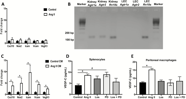
Figure 3.
Angiotensin II-activated immune cells induce LEC activation and inflammation: (a) Gene expression changes in LECs treated with vehicle (saline) or Ang II for 24 hours. (b) Gel electrophoresis of PCR products from mouse kidney (positive control) and primary mouse LECs amplified for Agtr1a and Agtr2. Rn18s was used as housekeeping gene. (c) Gene expression changes in LECs treated for 24 hours with conditioned media from saline-treated or Ang II-treated splenocytes. VEGF-C levels in conditioned media derived from (d) splenocytes or (e) peritoneal CD11b+ cells treated with saline, Ang II, Ang II + Los, Ang II + PD, or Ang II + Los + PD for 48 hours as measured by ELISA. All experiments were performed in triplicate. Results are expressed as mean ± SEM, and statistical analyses were performed with Student’s t test. #0.05 < P < 0.1 vs. Control; *P < 0.05 vs. Control or Control CM. Abbreviations: Ang II, angiotensin II; CM, conditioned media; LEC, lymphatic endothelial cell; PCR, polymerase chain reaction.

