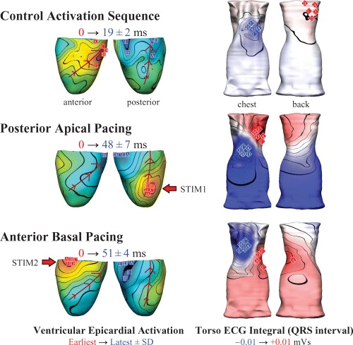
Figure 1.

Simultaneous epicardial (left) and body surface ECG (right) imaging in the anesthetized pig (n = 5) during sinus rhythm (control heart rate 156 ± 28 bpm; mean arterial blood pressure [ABP] 122/80 mm/Hg), and epicardial pacing (approximately 25 ppm above the intrinsic rate). Posterior apical pacing (STIM1) significantly increased the dispersion of epicardial activation from 19 ± 2 ms (control) to 48 ± 7 ms (p<0.01) and decreased the ABP by approximately 45/25 mmHg. Activation dispersion for antero‐basal epicardial pacing (STIM2) was 51 ± 4 ms (p<0.01 compared to control) and the ABP dropped by approximately 45/20 mmHg. Epicardial activation and torso ECG integral maps show averaged data, with individual observations indicated for earliest (red squares) and latest (blue squares) epicardial activation, and positive (red diamonds) and negative (blue diamonds) extrema of the torso ECG QRS integral maps. The minor nature of the variations in body surface activity suggests that some electrical features may be robustly obtainable using in vivo inverse ECG imaging.
