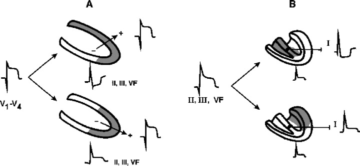
Figure 2.

(A) Shows how in the case of ST elevation in precordial leads as a consequence of occlusion in LAD, the ST changes in reciprocal leads (II, III, VF) allow to identify whether the occlusion is in proximal (above) or distal LAD (below), and (B) shows how in the case of ST elevation in II, III, and VF the changes of ST in other leads (lead I) give us information on if the inferoposterior wall infarct is due to RCA (above) or LCX (below) occlusion.
