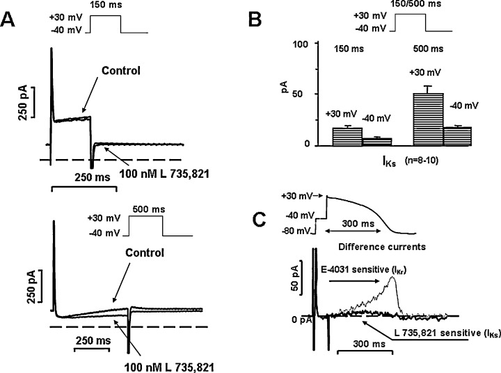
Figure 7.

Panel A. L‐735,821 sensitive (IKs) currents in human ventricular myocytes after application of short (150 ms, upper) and long (500 ms, bottom) depolarizing test pulses. Insets show applied voltage protocols. Panel B. Average IKs currents at the end of a short (150 ms) a long (500 ms) depolarizing test pulses to +30 mV, and peak tail currents at −40 mV. Panel C. L‐735,821 sensitive (IKs) difference current recorded during an “action‐potential‐like” test pulse in human ventricular myocytes in the absence of any sympathetic agonist. The dotted line shows superposed the E‐4031 sensitive (IKr) current measured in similar condition [modified from Ref. 59, used with permission].
