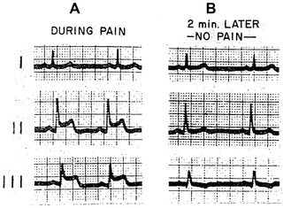
Figure 1.

(Case 1) Electrocardioram of variant type of angina pectoris. A, during spontaneous pain; note ST elevation in leads 2 and 3 and slight depression in lead 1. B, two minutes later pain disappeared and ECG reverted to normal. (Reproduced with permission from the American Journal of Medicine. [27:375‐388, 1959]
