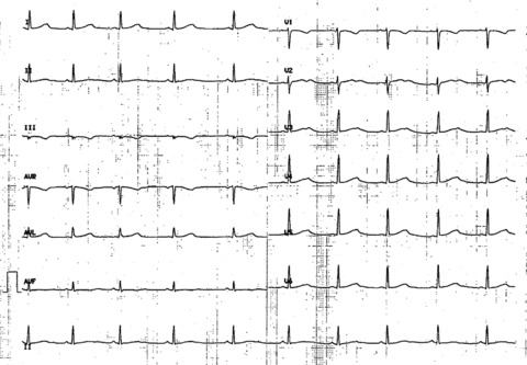
Figure 1.

12‐lead ECG recorded at admission. Sinus rhythm 58 bpm. PR: 140 ms. QRS: 80 ms, normal axis. Prolongation of QT interval with an obvious bifid T wave in precordial lead V2‐V3, and a subtle bifid T wave in V4‐V6. QTc: 560 ms.

12‐lead ECG recorded at admission. Sinus rhythm 58 bpm. PR: 140 ms. QRS: 80 ms, normal axis. Prolongation of QT interval with an obvious bifid T wave in precordial lead V2‐V3, and a subtle bifid T wave in V4‐V6. QTc: 560 ms.