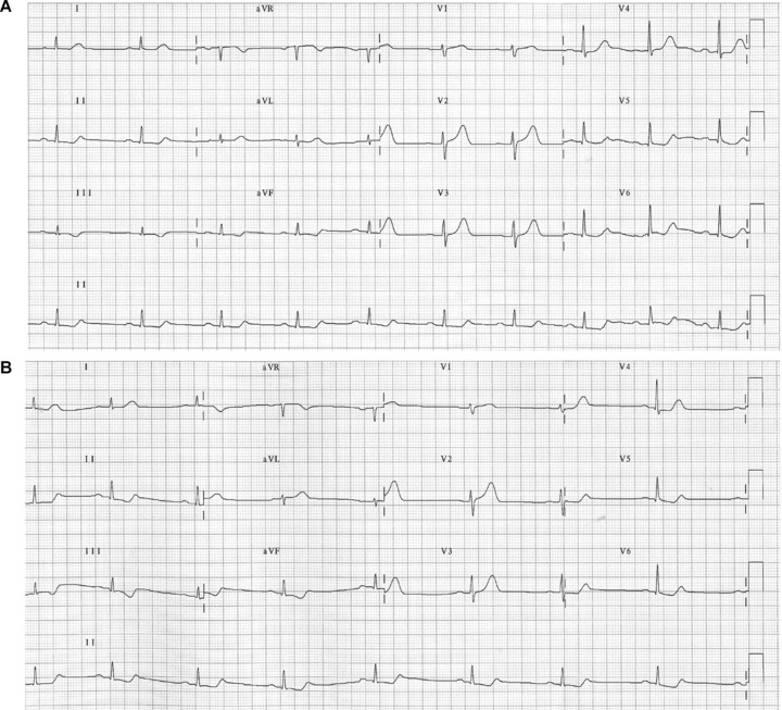
Figure 1.

(A) Electrocardiogram of a patient with acute coronary syndrome (ACS) due to coronary descending artery occlusion, obtained with standard electrode placement. (B) Electrocardiogram of the same patient obtained with Mason‐Likar system, showing markedly reduced R voltage in lead I and aVL, and increased in lead III and aVF. The reciprocal changes in inferior leads due to coronary descending artery occlusion are further accentuated on displacement of the limb electrodes to the torso.
