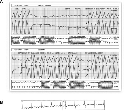
Figure 10.

(A) Healthy 35‐year‐old patient, with palpitations. Holter recording shows rapid bursts with wide complexes and morphology similar to that of ventricular flutter. Detailed examination of the recording allows identification of the basal rhythm (arrows) hidden among the wide artifactual complexes. (B) Patient with Parkinson's disease whose ECG simulates atrial flutter in DIII while V4 clearly shows P waves.
