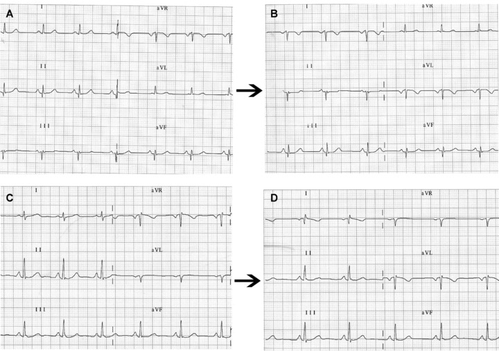
Figure 3.

(A) Frontal plane leads in a healthy patient with left shift of the QRS electrical axis. (B) on interchanging left arm and right arm electrodes, the ECG shows the classical pattern of P, QRS, and T as negative waves in lead DI and positive in aVR. However, in the vertical heart (C) the most striking finding may be (D) a QR or qR complex in lead DI with negative P wave or negative/positive in aVR.
