
Figure 5.
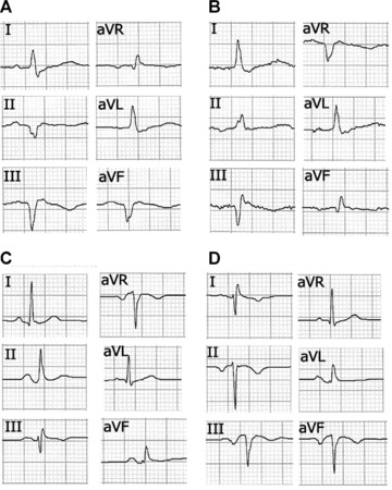
(A) Frontal plane leads in a 60‐year‐old male patient showing inferior myocardial infarction in chronic phase. (B) The same patient with right arm‐left leg electrode interchange. In this case, aVF resembles the usual aVR lead and II is inverted. The Q wave in lead II disappears and its depth in aVF is reduced, thus simulating less extension of the necrosis. (C) A 30‐year‐old woman without cardiopathy with correctly placed electrodes. (D) Tracing of the same patient with clockwise electrode rotation without ground electrode displacement, showing negative QS and P wave complexes similar to those obtained with right arm‐left leg electrode interchange obtained in a patient without previous inferior infarction.