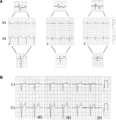
Figure 7.

(A1) Subject without cardiopathy and the V1 and V2 electrodes correctly placed; the P wave is positive in both leads. (A2) V1 and V2 electrodes placed on the 3rd intercostal space; the P wave is positive in both leads. (A3) rSr’ pattern preceeded by a negative P wave in V1 and V2; this suggests high placement of these electrodes (on the 2nd intercostal space). (B) Leads V1 and V2 in a patient without cardiopathy and normal ST segment, obtained with electrodes correctly placed in the 4th and 3rd intercostal spaces. If the electrodes are placed on the 2nd intercostals space, we see rectification and increased ST segment in V1 and V2.
