FIG 2.
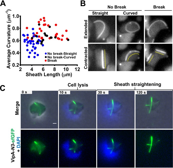
Straightening of T6SS sheath upon contraction and cell lysis. (A) Scatterplot showing the dispersion of the extended sheath length and the average curvature of contraction types in ampicillin-treated cells as follows: normal contraction, straightened (No break-Straight; n = 33); normal contraction, slightly curved (No break-Curved; n = 15); and sheaths that break into pieces after contraction (Break; n = 31). Each data point represents a sheath. (B) Sheaths formed in ampicillin-treated cells that contract normally (No Break) and straighten after contraction (left) or remain slightly curved (middle) and those that break (Break) into pieces after contraction (right). The dashed lines indicate the contracted sheath. Images were taken from a 5-min time-lapse video (10 s per frame). (C) Noncontractile VipA-N3–sfGFP curved sheath straightening after cell lysis induced by 80 μg/ml colistin–0.1% Triton X-100. Nucleic acid was stained with 10 μg/ml DAPI. Top row, merge of phase, GFP (green), and DAPI (blue) channels. Bottom row, green and blue channels only. The original video was an 8-min time-lapse video (10 s per frame). Scale bar, 1 μm. The full video is included in Movie S3 in the supplemental material (cell1). An additional example is provided in Fig. S3B in the supplemental material.

