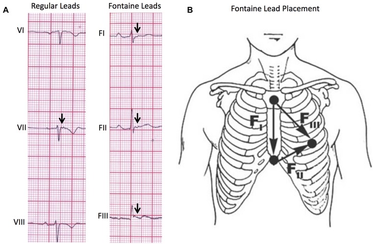
Figure 3.

(A) Comparison of regular lead placement versus Fontaine lead placement in the ability to detect epsilon waves (arrows). Using the Fontaine lead placement increases sensitivity of detecting epsilon waves so that they are detected in three leads (FI, FII, FIII) rather than one lead in the regular placement. (B) Fontaine bipolar precordial lead placement. In this modified technique, the ECG should be recorded at double speed (50 mm/s) and double voltage (20 mm/s) to improve the sensitivity for detection of epsilon waves.9
