
Figure 4.
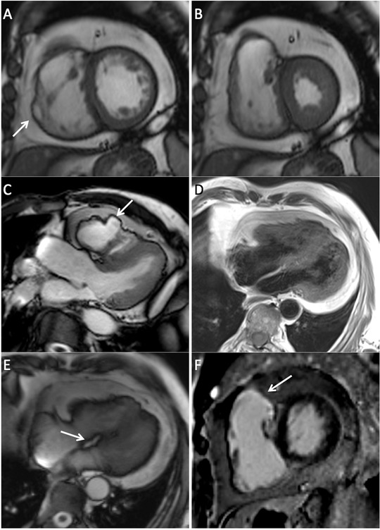
(A) Short‐axis bright blood SSFP image remarkable for abnormal amounts of epicardial fat, right ventricular wall thinning and aneurysm (arrow). (B) Short‐axis bright blood SSFP image in systole where right ventricular free wall thinning is demonstrated. (C) Left ventricular outflow tract SSFP image showing an irregular pattern of the right ventricular free wall with aneurysmal formation. (D) Double inversion recovery four‐chamber image demonstrating fat infiltration of the right ventricular free wall. (E) Four chamber SSFP image remarkable for the presence of fat infiltration in the basal portion of the interventricular septum (arrow). (F) Late gadolinium enhancement short‐axis image with presence of fibrosis in the right ventricular wall involving the outflow tract (arrow). The left ventricle is free of fibrosis.