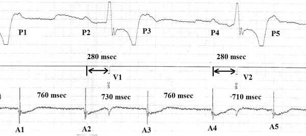
Figure 1.

Measurement of P‐P intervals during heart block. The upper channel, which is ECG Lead II, demonstrates five P waves, P1 to P5. There are two paced QRS complexes (V1 and V2) between the second and the third P and between the fourth and the fifth P waves. The middle channel, the marker channel, denotes the stimulus artifact of the paced QRS complexes. The bottom channel is the atrial electrogram, which demonstrates the five atrial events, A1 to A5, that correspond to the five surface P‐waves. The P2‐P3 interval shortening in the presence of a QRS (V1) was calculated by comparing A2‐A3 to A1‐A2. For this example, P‐P interval shortening = ((760–730)*100)/760 = 3.9%. According to our previous definition (2) VR is present in this instance.
