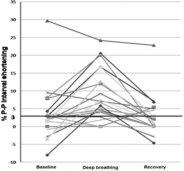
Figure 3.

The percent P‐P interval shortening surrounding QRS complexes during heart block in relation to deep breathing. Each line represents data from an individual patient, demonstrating the percent change in P‐P interval shortening surrounding QRS complexes at baseline (normal respiration), during deep breathing, and then during recovery. In 14 (70%) of these 20 patients the percent P‐P interval shortening was greatest during deep breathing.
