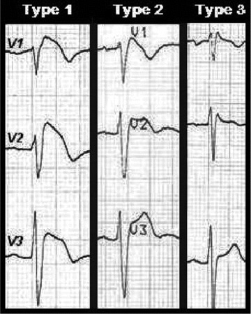
Figure 1.

In the type 1 ECG, the elevated ST segment (≥2 mm) descends with an upward convexity to an inverted T wave. This is referred to as the coved‐type Brugada pattern. The type 2 and 3 patterns have a saddle‐back ST‐T wave configuration, in which the elevated ST segment descends toward the baseline, then rises again to an upright or biphasic T wave. The ST segment is elevated ≥1 mm in type 2 and <1 mm in type 3.
