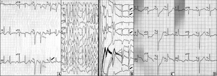
Figure 2.

The second exercise test and the resting ECGs on the other day. (A) During the exercise test, the J wave was initially seen in lead V6 (black arrow) with the upsloping ST‐segment depressions in V1–V4 (open arrows). (B) As the exercise test progressed, the polymorphic ventricular tachycardia occurred which was ceased by ICD shock—after the shock the J waves were observed in leads II, III, aVF, and V6 (black arrows). (C) The ECG during a resting chest pain, ST‐segment elevations in inferolateral leads with reciprocal changes in anterior leads can be seen. (D) Immediately after the nitroglycerin administration, the ST‐segment elevations (from panel C) and the chest pain disappeared.
