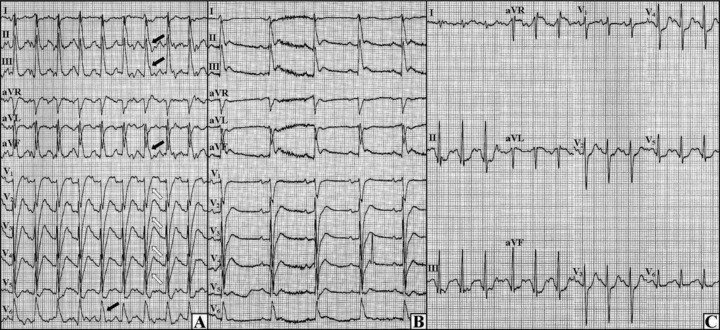
Figure 3.

The third and fourth exercise tests. (A) Final phase of the third test, the J waves in inferolateral leads (black arrows) with upsloping ST‐segment depressions in anterior leads (open arrows) can be seen. (B) The third test, the ECG during pulseless electrical activity is shown. (C) Final phase of the fourth test, no J wave can be observed.
