
Figure 1.
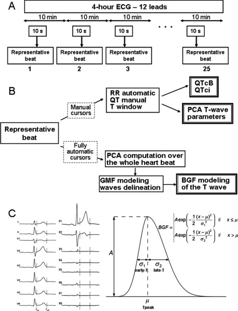
(A) Study design and time points for ECG extraction. (B) Biomarker categories: (1) manual cursors are set for both conventional ECG time intervals and markers derived from a PCA analysis on the T wave, (2) fully automatic biomarkers computation from the bi‐Gaussian function (BGF) model. (C) Left part: standard eight‐lead ECG change to PCA eight‐lead transformation. Right part: Bi‐Gaussian function model of the T wave.