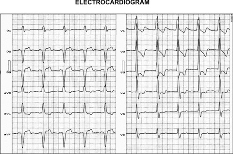
Figure 2.

(A) This ECG depicts extreme left QRS axis deviation (‐85°), SIII > SII, final S wave in left leads V5 and V6; all ECG features compatible with LAFB. PAF, R wave voltage “in crescendo” from leads V1 to V3 and decreasing from leads V4 to V6, small initial Q wave in leads V1 toV3, absence of initial Q wave in leads V5 and V6; all ECG features compatible with LSFB. The diagnosis is Left bifascicular block: LAFB + LSFB.
