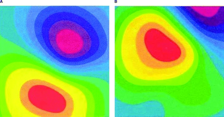
Figure 5.

Example for possible problem arising if sensor positioning is guided by chest wall anatomy. The cardiac magnetic field is correctly covered by magnetocardiographic sensors (left, a). The cardiac magnetic field is partially outside the magnetocardiographic area (right, b).
