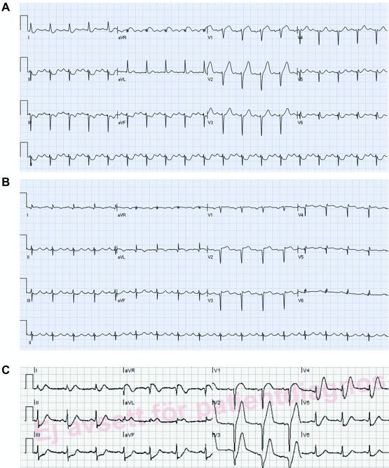
Figure 4.

(A) ECG of a 61‐year‐old female with severe chest pain. There is sinus tachycardia with ST elevation in aVR, ST depression in the inferior leads and mild upsloping ST depression in leads V3–V4 and tall T waves in V1–V3. Coronary angiography showed three vessel disease with total occlusion of the LAD. Left ventricular ejection fraction 20%. Patient underwent CABG. (B) Few days later, ECG shows QS wave in V2 with mild ST elevation in V2–V5, compatible with recent anterior STEMI. (C) An ECG of a patient with acute myocardial infarction. There is upsloping ST depression with positive T waves in leads I, II, III, aVF, V2–V6. There is ST elevation in leads aVR and V1.
