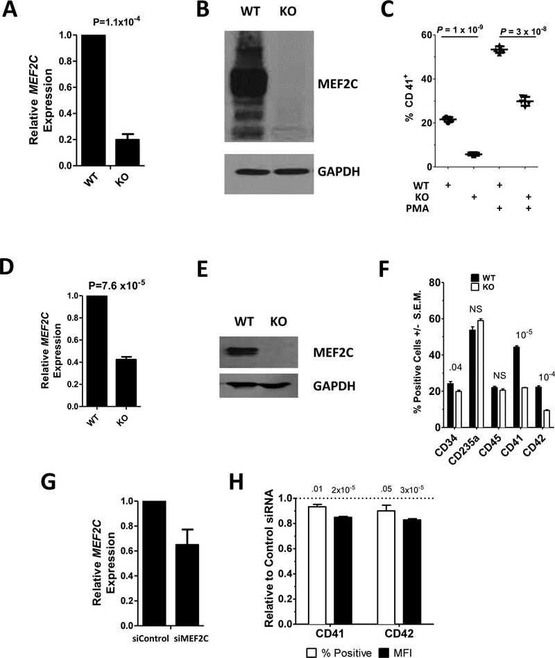
Fig. 1. MEF2C deficient cells are defective in megakaryocytic differentiation.
MEF2C (A,D,G) mRNA and (B,E) protein were assayed by qRT-PCR and Western Blot, respectively in: (A,B) Meg01 (WT) and Meg01-MEF2CKO (KO); (D,E) CHOP10 (WT) and CHOP10-MEF2CKO iPSCs; and (G) CB CD34+ derived megakaryocytes transfected with control (siControl) or MEF2C (siMEF2C) targeting siRNA. (C) Surface CD41 levels in Meg01 (WT) and Meg01-MEF2CKO (KO) cells were measured by flow cytometry cells before and after PMA treatment. (F) Percent of cells positive for surface marker expression in CHOP10 (WT) and CHOP10-MEF2CKO (KO) cells measured by flow cytometry. (H) Surface CD41 and CD42 percent positive and mean fluorescent intensity (MFI) of CB CD34+ derived megakaryocytes transfected with anti-MEF2C siRNA. Values are presented as relative to control siRNA transfected cells. P-value above bars. NS=non-significant.

