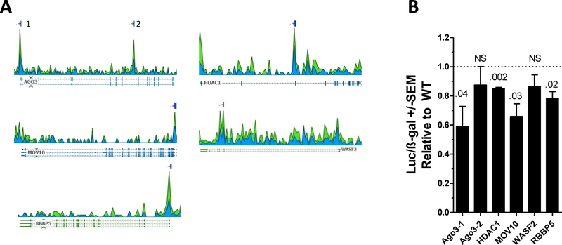
Fig. 3. Identification of MEF2-C responsive genomic sequences in target genes.
(A) Read depth of MEF2C ChIP-Seq in GM12878 B-cells. Reads are aligned either to the forward (blue) or reverse (green) strands. Lines above the coverage plot indicate peaks identified using the optimal Irreproducible Discovery Rate (IDR).23 The AGO3 gene contains two peaks, labeled 1 and 2. (B) Reporter constructs containing the putative MEF2C binding sites were transfected into WT and MEF2C KO cells and assayed for luciferase activity. Results are normalized to ß-gal activity. P-value above bars. NS=non-significant.

