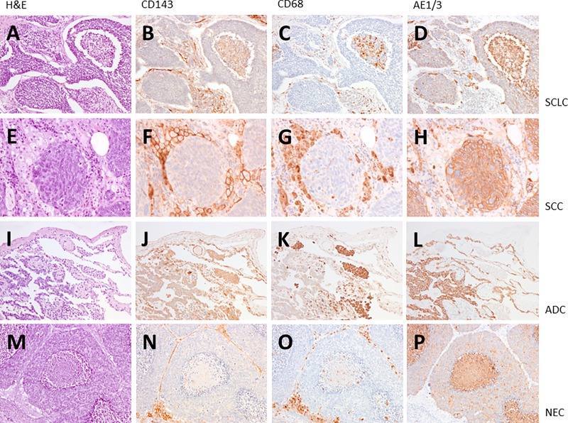
Fig 1. ACE expression in lung cancer.
We performed immunostaining on the lung cancer specimens with anti-ACE mAbs CG2. We have analyzed 3 cases of SCLC (small cell lung cancer; pictures A-D), 9 cases of NSCLC (non-small lung cancer), here 3 squamous cell carcinoma (SCC; pictures E-H), 3 adenocarcinoma (ADC; pictures I-L) and 3 neuroendocrine carcinoma (NEC; pictures M-P). ACE expression and localization is shown by brown color. Compared to anti-CD68 (macrophages; pictures C, G, K, O) and AE1/3 (cancer cells; pictures D, H, L, P) ACE is expressed strongly in macrophages and endothelial cells of the tumor vascularization in all types of lung cancer analyzed (pictures B, F, J, N). A weak expression of ACE in cancer cells was only found in adenocarcinoma (picture J). ACE was not detected in cancer cells of SCLC, SCC and NEC (pictures B, F, N). A, E, I, M: H&E; B, F, J, N: CD143 (ACE); C, G, K, O: CD68; D, H, L, P: AE1/3. Magnification x100 (A-D, I-L, M-P), x200 (E-H).

