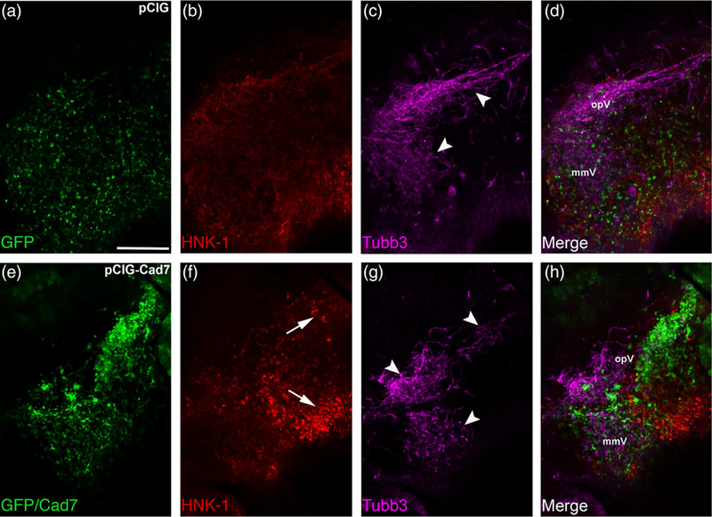
FIGURE 10.
Elevated levels of Cadherin-7 in migratory neural crest cells alter the gross morphology of the trigeminal ganglion. Representative lateral views (optical section) of the forming trigeminal ganglion in an HH15 chick head after electroporation of the pCIG control vector (pCIG, a–d) or the pCIG-Cadherin-7 vector (pCIG-Cad7, e–h) at the 3ss, followed by whole-mount immunohistochemistry for HNK-1 (red) and Tubb3 (purple). Merge images are shown in (d, h). A trigeminal ganglion electroporated with the control pCIG vector in neural crest cells (a) exhibits a bilobed morphology, with neural crest cells (b) coalescing with placodal neurons (c, arrowheads). A trigeminal ganglion electroporated with pCIG-Cad7 in the neural crest (e) possesses neural crest cells that migrate to the anlage, but these cells appear to aggregate together, thus altering their general distribution in the anlage (e, f, arrows). Placodal neurons are also affected, exhibiting a less compact appearance (g, arrowheads). Together, this leads to an abnormal ganglion shape relative to control. The ophthalmic (opV) and maxillomandibular (mmV) lobes of the trigeminal ganglion are indicated in (d) and (h). Scale bar in (a) is 200 μm and applies to all images

