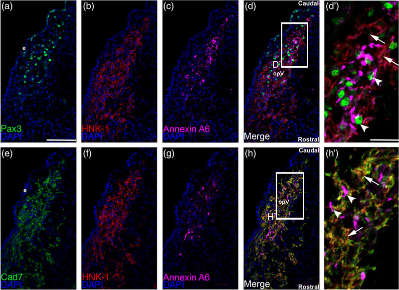
FIGURE 2.
Cadherin-7 protein localizes to HNK1-positive neural crest cells but not Pax3-positive placode cells or placodal neurons. Representative serial/adjacent transverse sections taken at the axial level of the forming ophthalmic lobe of the trigeminal ganglion at HH15 followed by triple-label immunohistochemistry for either Pax3 (a, green), HNK-1 (b, red), and Annexin A6 (c, purple), or Cadherin-7 (e, green), HNK-1 (f, red), and Annexin A6 (g, purple). (d′) Higher magnification image of the boxed region in (d) shows placode cells or placodal neurons (Pax-3- and Annexin A6-positive cells, arrowheads) that are not labeled with HNK-1 and thus are different from HNK-positive neural crest cells (arrows). (h′) Higher magnification image of the boxed region in (h) reveals Cadherin-7 immunoreactivity solely in HNK-1-positive neural crest cells (arrows), while Annexin A6-positive placodal neurons (arrowheads) are devoid of Cadherin-7. Rostral and caudal orientation and the ophthalmic lobe of the trigeminal (opV) are indicated in (d) and (h). DAPI (blue) labels cell nuclei. e = ectoderm. Scale bar in (a) is 67 μm and applicable to (b–h), while scale bar in (d′) is 20 μm and applicable to (h′)

