FIG. 5.
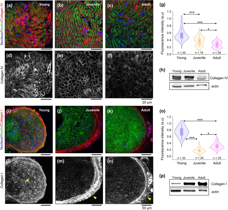
Changes in peripheral nerve ECM collagens during sciatic nerve development. Representative confocal microscopy images of sciatic nerve cross sections showing fluorescently labeled distribution of the basal membrane collagen type IV network (a)–(f) and collagen type I (i)–(n) in young, juvenile, and adult mice. Myelin and nuclei are stained in green (FluoroMyelin) and blue (DAPI), respectively. (g) and (o) Quantification of immunofluorescence for endoneurial collagen type IV and type I, respectively. Data are represented as violin plots with overlaid box-plots. Red dot = mean; box = 25th and 75th percentile. n indicates the number of areas analyzed from 2 different animals per developmental stage. Western blot shows decreased levels of collagen type IV with nerve maturation (h), whereas collagen type I levels increase with nerve maturation (p). * indicates a significant difference with p < 0.05 and *** with p < 0.0001 obtained using a Mann-Whitney test (g) and paired two-tailed t-test (o).

