Figure 1.

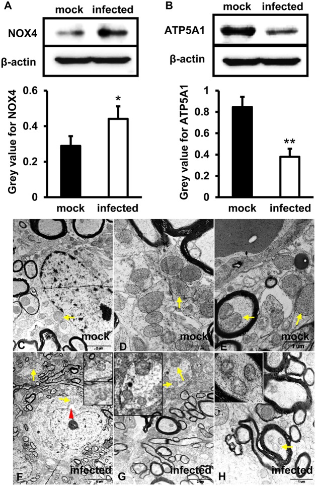
Mitochondrial dysfunction was related to an increased level of NOX4 and decreased expression of ATP5A1 in HEV infected brain tissues. (A,B) in vitro, HBMVECs were inoculated with 300 MOI HEV for 48 h for western blot, and HEV-negative homogenate served as control. Data showed that expression of NOX4 in HBMVECs treated with HEV for 48 h was significantly increased compared with mock group (*p < 0.05). Meanwhile, ATP5A1 was detected significantly attenuated in HEV inoculated cells (**p < 0.01). (C-H) In vivo, brain and spinal cord tissues that detected for HEV-RNA positive were selected for ultrastructural study. (C–E) Mitochondria were observed with clear cristae folded by the inner membrane in brain tissue of mock group. (F–H) Mitochondria with loss of cristae were found in tissues of HEV infected animals (arrows). The rough endoplasmic reticulum of neuron cells was also observed with mild distended cisternal space in HEV infected tissues (arrowhead). For western blot, gray value was analyzed with ImageJ to quantitatively analyze the expression levels of targeted proteins according to the level of exposure gray (resolution). Data were finally normalized to the expression of anti-β-actin.
