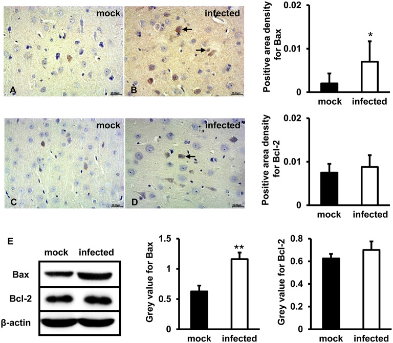
Figure 3.
Pro-apoptotic protein Bax but not Bcl-2 was upregulated following HEV infection. (A–D) HEV-RNA positive brain tissues collected on 14, 21, and 28 dpi were used for the immunohistochemistry study of Bax (rabbit polyclonal IgG) and Bcl-2(rabbit polyclonal IgG). Goat anti-rabbit IgG was chosen as secondary antibody. The positive signal was measured via the Motic Med 6.0 CMIAS Image Analysis System. Data showed that Bax was mainly distributed in cytosol of neuron cells, vascular endothelial cells and few microglial cells of HEV infection tissues with increased amount compared with mock group (*p < 0.05). Bcl-2 was detected in few neurons and vascular endothelial cells in both groups. (E) For western blot, HBMVECs were inoculated with 300 MOI HEV for 48 h and HEV-negative homogenate served as control. Data showed that expression level of Bax was significantly higher in HBMVECs inoculated with HEV (**p < 0.01), but induction of Bcl-2 was not significant. For western blot, gray value was analyzed with ImageJ to quantitatively analyze the expression levels of targeted proteins according to the level of exposure gray (resolution). Data were finally normalized to the expression of anti-β-actin.

