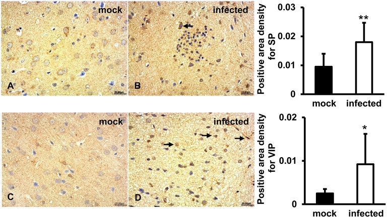
Figure 5.
Immunoregulation of the brain tissue during HEV infection. HEV-RNA positive brain tissues collected on 14, 21, and 28 dpi were used for immunohistochemistry study of SP (rabbit polyclonal IgG) and VIP (rabbit polyclonal IgG). Goat anti-rabbit IgG was chosen as secondary antibody. The positive signal was measured via the Motic Med 6.0 CMIAS Image Analysis System. (A,B) A positive signal of SP was found in the cytoplasm of neuronal cells of brain tissue in both groups and was significantly induced in HEV infected animals compared with animals from mock group (**p < 0.01). (C,D) Expression of VIP was observed in vascular endothelial cells, astrocytes and oligodendrocytes in both groups, with significantly increased levels in HEV infected tissues compared with tissues from mock group (*p < 0.05).

