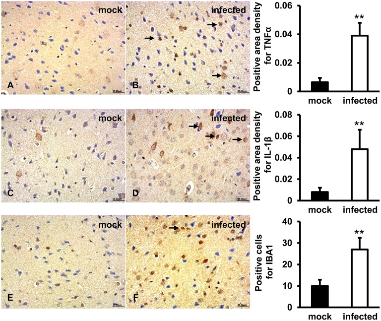
Figure 6.
Increased pro-inflammatory responses were examined in HEV infected animal brain tissues. HEV-RNA positive brain tissues collected on 14, 21, and 28 dpi were used for immunohistochemistry study of TNFα (rabbit polyclonal IgG), IL-1β (rabbit polyclonal IgG), and IBA1 (rabbit polyclonal IgG). Goat anti-rabbit IgG was chosen as secondary antibody. The positive signal was measured via the Motic Med 6.0 CMIAS Image Analysis System. (A,B) Positive signal of TNFα was diffusely observed in a large number of microglial cells, astrocytes and few pyramidal neurons in HEV infected animal brain tissues, and expression level was significantly induced compared with mock group (**p < 0.01). (C,D) The expression level of activated IL-1β was found in microglia, astrocytes, and few neuronal cytoplasms and was significantly increased in HEV infected brain sections compared with tissues from mock group (**p < 0.01). (E,F) Positive expression of IBA1 was detected in microglial cells with significantly elevated levels in brain tissues infected with HEV compared with mock group (**p < 0.01).

