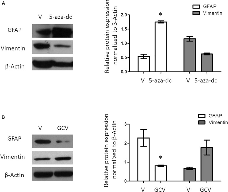
FIGURE 3.
Protein expression analysis of GFAP and Vimentin in GFAP-TK mouse spinal cord-derived neurospheres cultured in vitro. (A) GFAP-TK neurospheres cultured for 2 weeks were treated with vehicle (DMSO [V]) or 5-aza-dc (48 h). Analysis of protein expression (a) by Western blotting used specific antibodies against GFAP and Vimentin. Densitometry analysis of proteins was performed using the ImageJ software. Values were normalized to β-Actin in three independent experiments and results represented in the graphics. (B) GFAP-TK neurospheres cultured for 2 weeks were treated with vehicle (DMSO [V]) or GCV (24 h). Analysis of protein expression was quantified by Western blotting (a), and the corresponding quantification by densitometry is shown. ∗p < 0.05, determined by a Mann Whitney U test were considered statistically significant.

