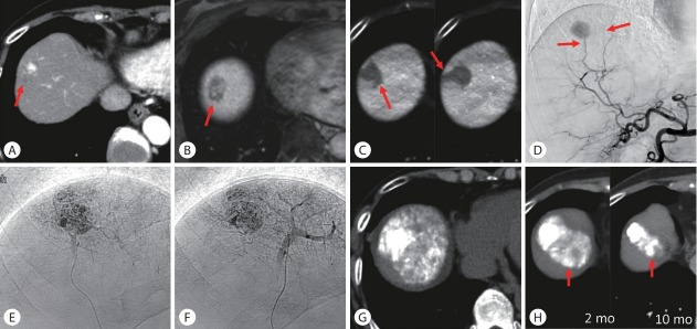
Figure 2.
Ultraselective conventional transarterial chemoembolization (cTACE) for hepatocellular carcinoma (HCC) with extracapsular and microvascular invasion. (A) Arterial phase computed tomography (CT) showed a hypervascular HCC with hypovascular extracapsular invasion (arrow) in segment 8. (B) Hepatobiliary-phase of gadoxetate disodium-enhanced magnetic resonance imaging also showed extracapsular invasion (arrow). (C) Cone-beam CT (CBCT) during arterial portography showed microvascular invasion around the tumor (arrows). (D) Celiac arteriogram showed tumor stain supplied by two feeders (arrows). (E, F) Each feeder was selectively embolized using a 1.5-F tip microcatheter and portal veins were markedly opacified during ultraselective cTACE. (G) Unenhanced CT performed 1 week after ultraselective cTACE showed dense iodized oil accumulation in the entire tumor including the hypovascular tumor portion. (H) Serial CT images showed that the tumor has remained well controlled 10 months after ultraselective cTACE. Iodized oil was also retained in the normal liver parenchyma (arrows), indicating hepatic necrosis. mo, months.

