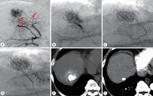
Figure 5.
The order of embolization of each feeder. (A) Arteriogram of the anterior segmental artery of the right hepatic artery showed a tumor supplied by three tumor-feeders (arrows). (B-D) Each tumor-feeder was selectively embolized from distal to proximal to avoid inadvertently occluding the distal tumor-feeders with overflowing embolic agents. (E) Unenhanced computed tomography (CT) performed 1 week after ultraselective conventional transarterial chemoembolization (cTACE) showed dense iodized oil accumulation in the tumor. (F) Arterial phase CT performed 10 years and 4 months after ultraselective cTACE showed that the tumor has remained well controlled.

