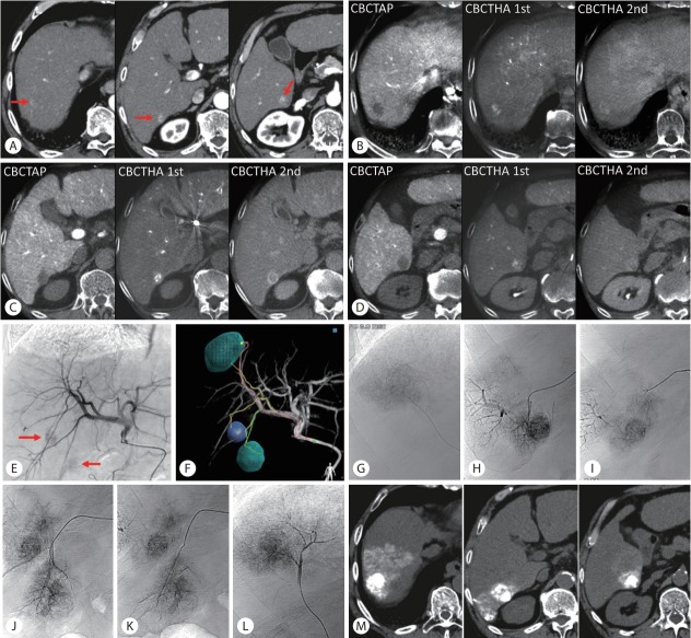
Figure 6.
Cone-beam computed tomography (CBCT) and transarterial chemoembolization (TACE) guidance software. (A) Arterial phase computed tomography (CT) showed 3 small hepatocellular carcinomas in the right hepatic lobe (arrows). (B-D) CBCT during arterial portography (CBCTAP) and dual-phase CBCT during hepatic arteriography (CBCTHA) could depict all tumors. (E) Common hepatic arteriogram showed two faint tumor stains (arrows). However, the tumor-feeders were unclear. (F) TACE guidance software identified the feeders of each tumor. (G-L) Each tumor-feeder was selectively embolized. One branch (Fig. 6I) was not detected by TACE guidance software, but it was embolized on our decision. (M) Unenhanced CT performed 1 week after ultraselective conventional TACE showed dense iodized oil accumulation in all tumors with a sufficient safety margin.

