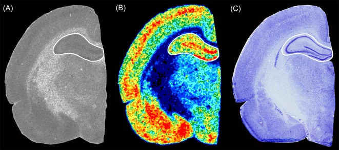
Figure 1.
Exemplary section from a P20 rat brain showing the hippocampal level of (A) an autoradiographic image of [3H]SR 95531 binding to GABAA receptors, (B) the respective color-coded image of the same autoradiographic image, and (C) an adjacent Nissl stained section. The Paxinos and Watson rat brain atlas (Paxinos and Watson, 2005) was used to verify the regions of interest.

