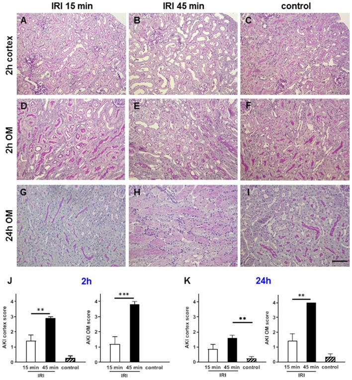
Figure 3.
Renal morphology at two and 24 h after IRI. PAS staining revealed that mild AKI was present already after 2 h in short term IRI (15 min, A,D,G,J). Longer ischemia time of 45 min caused more severe AKI mainly in the outer medulla (B,E,H,K). Sham kidneys served as controls and showed normal renal morphology (C,F,I). Signs of AKI were less at 24 h after IRI in this short IRI group and more pronounced in the prolonged IRI group (G–I, mean ± SEM, **p < 0.01, ***p < 0.001, bar: 100 μm, n = 6 mice/group, one way ANOVA).

