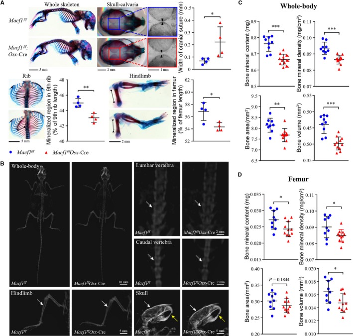
Figure 2.

Deficiency of Macf1 delayed bone ossification and decreased bone mass. (A) Alizarin red and Alcian blue staining of whole‐mount skeletal of newborn Macf1f/f and Macf1f/fOsx‐Cre mice (n = 4 per group). Bone ossification of skull, rib and hindlimb was quantified by width of cranial suture, mineralized region rate of 9th rib and mineralized region rate of femur, respectively. (B) DXA analysis of hindlimb, lumbar vertebra, caudal vertebra and skull from 3‐month‐old Macf1f/f and Macf1f/fOsx‐Cre mice. The radiodensity of hindlimb, lumbar vertebra, caudal vertebra and skull was indicated by white arrows. (C, D) Quantification of bone mineral content, bone mineral density, bone area and bone volume in whole body (C) and femur (D) from Macf1f/f and Macf1f/fOsx‐Cre mice (n = 9 for Macf1f/f, n = 11 for Macf1f/fOsx‐Cre). Data are presented as means ± SEM. *P < .05, **P < .01 and ***P < .001
