
Figure 7.
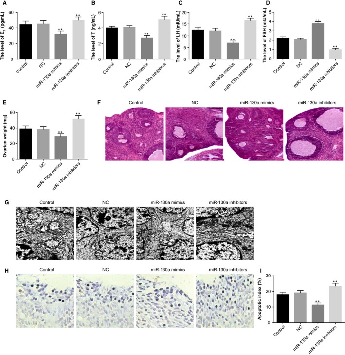
The effect of miR‐130a on the endocrine and ovarian granulosa cells in PCOS rat models. Note: A, serum levels of E2 in PCOS rat models determined by ELISA; B, serum levels of T in PCOS rat models determined by ELISA; C, serum levels of LH in PCOS rat models determined by ELISA; D, serum levels of FSH in PCOS rat models determined by ELISA; E, weight of ovaries of PCOS rat models; F, HE staining to observe histopathological changes of ovarian tissues of PCOS rat models (×100); G, the ultrastructures of ovaries of PCOS rat models observed under a TEM (×10 000); H, TUNEL staining to observe the apoptosis of ovarian granulosa cells in PCOS rat models (×200); I, quantitative analysis for the apoptotic index of ovarian granulosa cells in PCOS rat models; **P < .01 compared with the control group; PCOS, polycystic ovaries syndrome; ELISA, enzyme‐linked immunosorbent assay; E2, oestradiol; T, teststerone; LH, luteinising hormone; FSH, follicle‐stimulating hormone; TEM, transmission electron microscope; and TUNEL, terminal deoxynucleotidyl transferase‐mediated dUTP nick‐end labelling