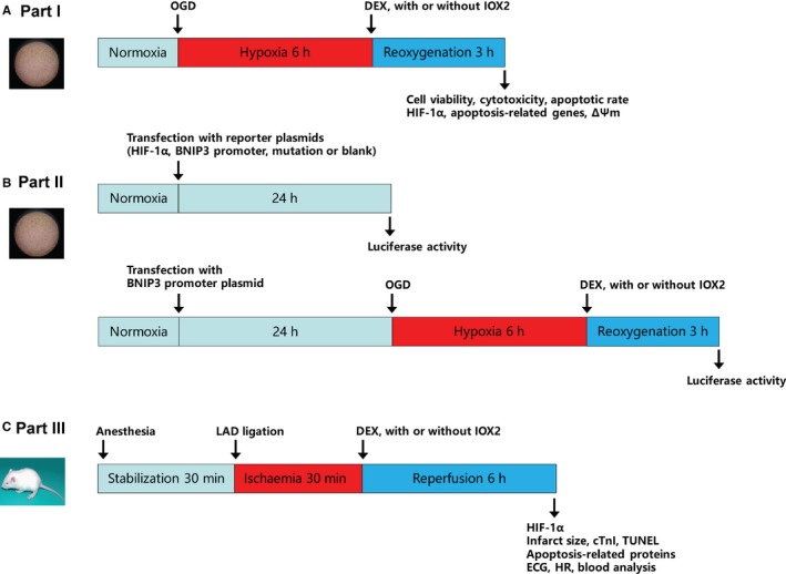
Figure 1.

Experimental protocols. A, Part I: neonatal rat cardiomyocytes were subjected to hypoxia/reoxygenation. B, Part II: cells were transfected with reporter plasmids and luciferase activity was assessed. C, Part III: rats underwent myocardial ischaemia/reperfusion. OGD, oxygen‐glucose deprivation; DEX, dexmedetomidine; LAD, left anterior descending; ∆Ψm, mitochondrial membrane potential; ECG, electrocardiography; cTnI, serum cardiac troponin I; and HR, heart rate
